


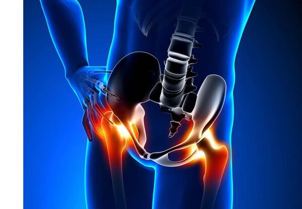
Thigh joint arthrosis (coxarthrosis)- It is a chronic degenerative joint disease that causes bone tissue deformity.With Coksartrosis, all components of the joint are involved in the pathological process: articular cartilage, cartilage bone structures, synovial shell, ligaments, capsules and surrounding muscles.In the case of the disease, the articular cartilage is destroyed, the micro-rimites of the bones and osteophytes appear, and inflammation of the ligament apparatus of the thigh joints.
In the world, all fifth people complain of joint problems with funds.It can be both pain, or limitation of movement in the joints, as well as a combination of these symptoms.Every second outpatient vision falls in patients with bone muscle disorders, and 66 % of cases are 65 years old.According to the latest epidemiological study, the prevalence of knee and thigh joint arthrosis in the adult population is 13 %.
Risk factors for the development of coxarthrosis:
- Genetic predisposition.A common cause of coccaartosis of the thigh joints is a congenital or acquired mutation type II type II prolagen type.
- Older age.The alleged cause of arthrosis prevalence in old age is a discrepancy between the external environment joint cartilage and its recovery capabilities.
- Flooring.Women experience osteoarthritis more often than men.This is due to the influence of estrogen female sex hormones on bone mineral metabolism.However, the influence of the floor is ambiguous - some authors believe, unlike other joint damage, there are no differences in the sexual basis of Cocsartus: in men, arthrosis of the thigh joint, as is often found in women.
- Excess body weight.The relationship is confirmed between excess body mass and arthrosis.Excess adhesive fabric increases the harmful load on the cartilage.In addition, fatty tissue produces pro -inflimatic enzymes that damage the cartilage tissue.
- Frequent development of bones and joints.According to studies, 80 %of coxarthrosis, which occurs for an obvious cause, is associated with previously diagnosed defects, in the development of the thigh joint - dysplasia and sublux.
- Hard physical labor.Excess load on the thigh joints with certain types of physical labor can be caused by joint damage and arthrosis.Agricultural workers, trenches, and people of similar work specialties are at risk.
- Injuries.The risk of developing coxarthrosis increases after the thigh joint is damaged.Moreover, both affected joints and both can be involved in this process.
- Professional gaming sport.Professional sports can cause coxarthrosis, both due to excessive loading on the joints and due to injuries.Potentially dangerous sports include heavy athletics, athletics jumping, parachute sports.
- Bones and joint diseases- Rheumatoid arthritis, psoriasis arthritis, joint infections, avascular necrosis, gout arthritis, etc.
- Endocrine pathologies- Hypothyroidism, hypoparathyroidism, acromegaly (impaired function of the anterior pituitary gland), diabetes, obesity.
If such symptoms are detected, consult your doctor.Don't be self -sufficient - it's dangerous to your health!
Symptoms of arthrosis of the thigh joints
The main symptoms of coxarthrosis are: pain, mobility restrictions and joints, their deformity, functional reduction of the lower limb and periodic swelling in the joints.
Pain of different intensity.Pain in the joint is initially minor and arises for a short time.They appear or are intense while walking or with other physical efforts, such as squares, inclinations and weight gain.As the disease develops, the pain is intense and even long rest does not bring relief.In addition, the pain is found in prolonged immobility and in one condition of the joint.
Patients complain about the so -called"Starting" pains in the thigh joints after sleeping, driving in the car and prolonging other."Start" for coxarthrosis lasts no more than 30 minutes.Pain is intense during hypothermia or in a stressful situation.They can be localized in the area of the buttocks or gums, on the anterior or lateral surface of the thigh.With the spread of lumbar plexus nerves, it can be moved from the center of the body or the thighs to the knee.Sometimes the pain extends to the lumbosacral spine and tail bone.
Restriction of joint mobility.The movements in the thigh joint with cocsartrose are limited due to pain.At the same time, rotation (returns both inside and out) and bringing the lower limb (movement in the middle of the body) is more often concerned, but may be limited (movement from the middle axis of the body), as well as flexibility and extension.Due to pain syndrome, the inability to make passive movements in the joint causes bias of the compensatory pelvis.The patient's gait is changing, the buttocks are back, the body deviates when the injured side of the weight is transferred.With bilateral injury, in patients with cocsartrosis, "duck gait" is created.
Coxarthrosis occurs periodicallySwelling in the jointWhich can be invisible because of the layer of muscle and fat.Also, the disease is characteristicDuring movement in the joints the crystal, their gradual deformation and the functional reduction of the lower limb.
Often, one joint is affected by the disease, then the process extends to others.But sometimes arthrosis has several joints at the same time and becomes polosostotarritis.Polyosteoarthritis is characteristic of older people, or hereditary predisposition and concomitant diseases - diseases of the bones, joints and endocrine disorders.
Pathogenesis of arthrosis of the thigh joints
In the pathogenesis of the thigh joints, mechanical injury (injuries and microtraumas due to physical force on the joint) and genetic, hormonal and metabolic factors play an important role.It is often impossible to determine which factor has affected the development of the disease in a particular patient, but often the disease develops with mechanical damage after tissue damage.
Tissue damage stimulates the division of cartilage tissue cells (chondrocytes), and the production of pro -infusion cytokines, which are usually found in the cartilage only in small quantities.Cytokines begin with an inflammatory process, for example, under the influence of anti-inflammatory cytokine IL-1, with enzymes that destroy the cartilage of the joint.Also, under the influence of cytokines, the production of TSOG-2 enzymes and other substances that have toxic effects on cartilage.
Synovites also play a major role in the development of coxarthrosis - inflammatory diseases of the joints or ligaments by accumulating fluid in the cavity.
Decreased elasticity and strength of articular cartilage associated with metabolic disorders results in a decrease in its resistance to mechanical stress.With coxartosis, all components of the joints participate in the pathological process, including subcondial bone.Since the large joints of the lower extremities are large in the body, they experience significant mechanical stress, causing mycoromas to be found in the subconditional plate and cartilage.As a result of the microbes, the subcondial bone is compressed, causing the regional growth of bone tissue - osteophytes.This, in turn, stimulates subsequent degradation of articular cartilage.
In some cases, arthrosis of the thigh joint is inherited.Hereditary arthrosis is likely to be a polygenic heritage - due to the action of many genes, each of which is weakly affected.Some diseases are the cause of mutation in genes that encode the articular cartilage macromolecules, which causes its decomposition.The genes responsible for dividing chondrocytes may also be affected.In addition, metabolic disorders are hereditary, for example, pyrophosphate arthropathy - a disease in which calcium pyrophosphate crystals accumulate in articular cartilage and synovial fluid.
Classification and stages of development of arthrosis of the thigh joints
For the causes of the disease, coxarthrosis is divided into two main forms: primary (idiopathic) and secondary (arising or due to other diseases).
Primary CoksarTrosis:
- Localized (only thigh joints operate):
- Unilateral;
- Mutual.
- Generalized (polyosteoarthritis) at least three joint group injuries (eg, thigh, knee and small joints or legs).
Used Arthrosis:
- Post -traumatic:
- Acute - as a result of severe injury;
- Chronic - due to some sports classes or as a result of professional activities.
- Metabolic diseases (Oconosis, Hemochromatosis, Wilson's Disease, Water Disease).
- Congenital pathologies and developmental defects (congenital thigh dysplasia, peresis disease, thigh epiphany, hypermobia syndrome, lower limb reduction, scoliosis, bone dysplasia).
- Endocrine pathologies (acromegaly, hypothyroidism, diabetes, hyperparathyroidism, obesity).
- Calcium salts (pyrophosphate arthropathy, calculation of tendonitis).
- Diseases of the bones and joints (rheumatoid arthritis, psoriasis arthritis, pedhetic disease, avascular necrosis, infections).
According to clinical manifestations, the following forms of coxarthrosis are distinguished:
- A little symptomatic.
- Manifestation, manifested by bright clinical symptoms:
- Rapidly progressive, in which symptoms develop within the first four years of the onset of the disease;
- Slowly progressive - clinically significant symptoms appear after five years of the course of the disease.
X -Ray picture may be detected by the arthrosis of the thigh joints:
- Hypertrophic - Increased paid response (injuries have been replaced by new tissue, for example, osteophytes appear);
- Atrophic (decreased tissue volume).
The stages of the disease can be radiologically and clinically determined.The most commonly used classification of Kelgren and Lawrence (1957) (1957) is the most commonly used classification of the thigh joint arthrosis.
Arthrosis stages in the X -ray classification
| Bact | Signs |
|---|---|
| 0 | There are no signs of arthrosis in x -ray pictures |
| 1 | The joint gap does not change, visualized individual regional osteophytes |
| 2 | The joint gap has not changed, important regional osteophytes are visualized |
| 3 | The joint gap height is moderately reduced, visualized by important regional osteophytes |
| 4 | The joint gap height is significantly reduced, visualized with significant regional osteophytes and subcondial osteosclerosis (bone tissue compaction under the lower surface of the cartilage) |
Classification (1961) is used to determine the clinical stage of the disease, which uses both clinical signs and visualization criteria.
Clinical stages of arthrosis
| Bact | Signs |
|---|---|
| 0 | The articular gap is unambiguous and unevenly narrow, the edges of the articular cracks are slightly marked (from osteophytes), a slight restriction of movements is marked |
| 1 | The articular gap is significantly narrow (50-60 %), significant osteophytes, subcondial osteoclamorosis, and cystic enlightenment in bone epiphysis;The clinic is predominantly limited by the limitation of mobility in the joints, during coarse crunchy movements, minor or moderate muscle atrophy |
| 2 | Deformation, joint strength;The articular gap is narrowed by more than 60-70 % of the norm or completely non -existent, with extensive osteophytes, subcondial cysts, articular "mice" are visualized bone, cartilage or mixed pathological formations located in the joint cavity. |
Complications of arthrosis of the thigh joints
With coxarosis, all complications are associated with pathological changes in the joints.
Coksartrosis course can be complicated by local inflammatory processes:
- Bursite - inflammation of the synovial bags in the joints;
- Tendovaginitis - inflammation of the vaginal membrane of the vagina of the muscle tendon;
- Nerve tunnel syndrome-pinches due to the formation of large osteophytes or deformation of the joint.
With the progression of coxarthrosis and its transition at the clinical stage II and III, the pain restricts the mobility of the joint, and over time, the joint anchorosis (fiber, bone or cartilage) occurs, accompanied by its complete immobility.
Joint significant deformation may causeBone fractures or aseptic necrosis.For Coksartrosis, aseptic necrosis of the thigh head is the strongest complication.
A pronounced coksartrosis can occurJoint subluxation and dislocationAlso penetrate the woman's head into the pelvic cavity.Desplanting and sublucing of the thigh joint causes pain (primarily severe, then boring and aching), during robbery and other physical efforts, as well as deformity of the joint, lame and sometimes affected limb.
Despite the lack of systemic manifestations of arthrosis itself, in modern clinical practice, more attention is paid to related diseases.These are abnormal conditions that exist or arise in the light of an existing disease.With regard to inflammatory reactions occurring during arthrosis, the formation of atherosclerotic boards on the inner walls of the vessels is improved, which increases the riskCardiovascular diseases.Depreciation of physical activity due to pain and joint mobilityObesity, depression and deterioration of quality of life.By prolonging non -anti -anti -anti -anti -anti -anti -anti -anti -anti -anti -anti -anti -anti -anti -anti -anti -anti -anti -anti -anti -antiUpper gastrointestinal sections are affected,And alsoThe risk of cardiovascular pathologies and kidney disease increases.
Diagnosis of arthrosis of the thigh joints
"Cocksarthrosis" is diagnosed on the basis of clinical manifestations and radiological examination.There are no characteristic laboratory signs for the diagnosis of arthrosis.
Between clinical manifestationsFor the diagnosis of thigh joint arthrosis, the main thing is pain and its character.Pain for the thigh joint arthrosis is found and gradually increases for several years (sometimes a few months in a rapidly progressive form).Pain is found or exacerbated by physical effort or permanent condition.If the patient begins to pain alone, then inflammation (synovitis) joins.The statement is marked up to 30 minutes in the morning and prolonged immobility.
Restriction of joint mobility gradually increases, with both active and passive movements.With the development of the disease, the joints are deformed, functional reduction of the length of the limb.
For a physical examinationThere is limitation of joint mobility, their deformity, reduction of limbs, pain in the joint palpation and large thigh spin, muscle atrophy.
Laboratory methodsNo need for the diagnosis of arthrosis of the thigh joints.However, they can be used for arthritis for the differential diagnosis of coxarthrosis (rheumatoid and chronic), as there are no inflammatory changes in the blood test and rheumatoid factor, and uric acid levels do not increase.In addition, using laboratory tests, contraindications are manifested for drug treatment methods.
Instrumental methodsFor the diagnosis of arthrosis of the thigh joints:
- Radiograph- This is the main method of diagnosis of thigh joint arthrosis.Radiography determines the characteristic changes of cocsartrosis: narrowing of the joint, osteophytes, cartilage, osteophytes and ulcer, subconcoral cysts and osteosclerosis.X -Ray examination is a classic method to determine the diagnosis of coxarthrosis, and radiological signs are based on classification of coxarthrosis.However, other methods of visualization of the joint are increasingly used, such as ultrasound and magnetic resonance imaging.
- Ultrasound examination (ultrasound) -The advantage of ultrasound is in the absence of radial load on the body.
- Magnetic -Raisonnesian Tomography (MRI)- Compared to other methods, this allows you to make joint damage more clearly.
- Arthroscopy—TheAllows you to determine the damage to the articular cartilage: from the zones of chondromation (softening of articular cartilage) less than 10 mm in diameter, to deep cracks that penetrate the sub -bone and produce deep ulcers.Surface and medium cracks and surface erosion can also be visualized.
Identifying Coksartrosis is usually not particularly difficult, but when evaluating a specific clinical situation, it is important to remember the possible secondary origin of the thigh joints (as with other diseases, such as endocrine disorders).
Treatment of arthrosis of the thigh joints
Treatment of arthrosis of the thigh joints can be both conservative (medicines, as well as non -operative) or operative.Conservative treatment is used in 1-2 stages of the disease, surgical-3 stage.Surgical treatment may be recommended in 2 stages, with persistent pain and absence of a reaction to conservative therapy.
Goals of Conservative Therapy:
- Improving quality of life - Reduce pain and increase joint mobility;
- Stop or slow down the development of the disease.
Unwanted treatment methods include:
- Unloading of thigh joints (body weight loss, creating additional support and transferring body weight to cane or gum);
- Physical education of physiotherapy;
- Methods of physiotherapeutic treatment.
Coxarthrosis treatment begins with non -tape methods, an important role in physiotherapy exercises.With severe pain, the patient should use support.With pronounced disease and contraindications in endoprosthetics, support should be used for life.
Kutsartosis Healing TherapyIncludes medications that reduce the symptoms of the disease.These are analgesics, as well as drug group drugs that have non -anti -anti -anti -drug (Nonsteroidal).NSAIDs are divided into area and selective.
Analgesics and NSAIDs are used to relieve pain and inflammation for a short time to relieve pain and inflammation.Currently, there is no approved advantage of one unwanted antiniflemian agent to another, so the choice of a particular drug depends on the side effects and the specific clinical situation caused by it.
It should be remembered that NSAIDs have many side effects.When taking them, the mucous membrane of the stomach and duodenum is affected, resulting in ulcers and bleeding.Many NSAIDs have toxic effects on the liver and kidneys.In addition, NSAIDs disrupt platelet aggregation and, as a result, the patient is thrown out of thrombosis and there is a tendency for bleeding.NSAIDs, which have prolonged use, inhibit hematopoiesis processes and can cause aplastic anemia and agranulocytosis.Taking selective NSAIDs causes significantly less complications.
Locally used ointments and gels cause less side effects than oral products.Medicines that have warming and pain reduction are used to treat arthrosis.They can contain turpentine, menthol, nicotinic acid esters, salicylates, bee venom.Also, NSAIDs have a good effect.
In the absence of analgesic and non -steroidal effects, or if it is not possible to choose the optimal dose of the drug, central action painkillers may be determined short -term.
In the case of inflammation, intraicular intake of corticosteroids is used.Corticosteroids are used no more than 2-3 times a year, as more frequent use can cause cartilage degeneration.
Slowly, medications that weaken the symptoms of the disease, include chondroprotectors, avocado or soy, inappropriate compounds of hyaluronic acid.These medicines are included in the European Anti -Rematic League recommendations to treat thigh joints arthrosis.Preparations reduce pain and improve joint mobility.
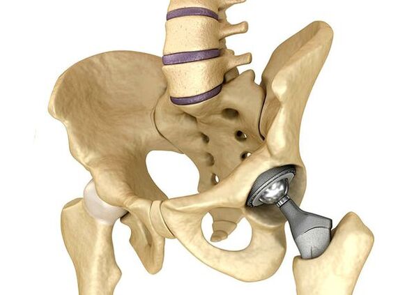
The endoprometry of the thigh jointsIt is used in severe cases of stage III when pain syndrome cannot be eliminated and joint mobility is significantly limited.The thigh joint prosthesis leads to a decrease in pain syndrome, improved joint function, and quality of life of the patient.The effect is maintained for 10-15 years, after which a second surgery may be required.During surgery, the thigh joint is replaced by artificial imitation of ceramic, metal (most commonly used titanium prostheses) or polymer.
Prognosis.Prevention
The prognosis of the thigh joint arthrosis is favorable to the patient's life, but the disease often causes disability.According to the World Health Organization, 80 % of older patients with coxarthrosis have mobility disorders, and 25 % cannot do daily affairs.In this regard, the main prevention of thigh joint arthrosis is important.
Prevention measures:
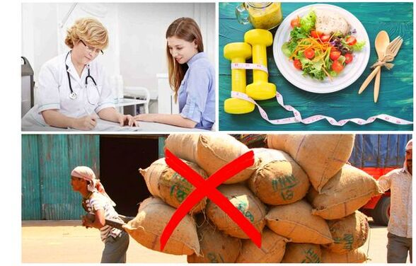
- Reducing body weight.Nutrition adjustment is necessary to reduce weight and load on the joint.In addition, the decrease in the volume of fatty tissue reduces the number of mediators released by it.
- Avoid heavy physical labor and sports overload.Physical overload is often the cause of arthrosis of the thigh joints, and moderate physical activity, on the contrary, improves the condition of the articular cartilage, maintains normal mobility and reduces load on other joints.
- Fix the underlying disease.If the patient is detected in diseases that can cause secondary coccarthrosis (endocrine, rheumatic, etc.), a major disease is necessary.Normalization of hormonal background and continuous remission of rheumatic diseases is the primary prevention of arthrosis and allows you to slow down its development.
- Find out a healthy lifestyle.A balanced diet with sufficient content of plants and animal protein, restriction of polyunist fatty acids and simple carbohydrates, as well as moderate physical activity, avoid coxarthrosis, even in the presence of risk factors.
Currently, the prevention of thigh joint diseases is mandatory in neonatology and pediatrics.Over time, congenital dysplasia regulated the thigh joint significantly reduces the risk of coxarosis in adulthood.